Medical drawings
Published in Features, Issue 4 (July/August 2018), Volume 26William Wallace Papers, Royal College of Surgeons in Ireland
By Fiona Fitzsimons

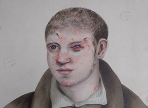
Above: ‘Peter Keating aged 24 years, skin lesions’, April 1838 (after 1895 described as granuloma). (RCSI/IP/Wallace/2/1/16/20)
One of the most accessible collections, the William Wallace Papers, is held in the archives of the Royal College of Surgeons in Ireland (RCSI). The Wallace collection comprises approximately 200 drawings in pen and ink, pencil and watercolour on paper. The drawings are accompanied by twelve casebooks, from 1810 to 1879. There are many more case-studies than there are drawings. It’s probable that the decision to make a visual record was based on how graphic the symptoms were. The collection was probably originally compiled as a learning/research aid for medics with an interest in skin and venereal diseases.
The drawings can be divided roughly between portraits and anatomical studies. The portraits are predominantly head and shoulders, with some half- and some full-length. These are candid portraits, with no attempt to soften or idealise the sitter. The artists’ commission was to show the disease, but as they drew from nature they revealed the patients/sitters as distinct characters. In all pictures the mood is sombre, and the patients appear anxious and downcast. In some drawings the patients’ clothes are evident, unwrapping yet another layer of the story. A woman’s white cap framing her lined face; a gentleman’s linen shirt fastened high on the collar-bone by a silk cravat; the wide blue and white stripe of a man’s flannel shirt, open to the waist—there’s a social history woven into our clothing.
The drawings of anatomical studies focus on specific body parts: a rash on the palm of a patient’s hand; the inside of a blistered gum; a tumour on the lip; skin folds on the neck or trunk; an abscess in the soft flesh of an armpit; an open wound on the thin skin of the shin; oozing pustules on the groin; ulcers on the penis and scrotum. The archival catalogue warns that ‘illustrations include graphic images of sexual organs’.
All the drawings record only the most basic evidence: the patient’s name, the date on which the drawing was made and, most importantly, the corresponding casebook volume and page number. The casebooks are almost artefacts in themselves, half-bound and hard-cover (32cm x 10cm), written up in a fine spidery hand. Each casebook has an index of patients’ names, diseases and the relevant page numbers: ‘John Gay, Throat, Penis, Skin; William Tindal: Penis, Groin; William Kearns: Testicle, Urethra …’. The names were set down as patients presented themselves, and there is no attempt to impose any alphabetical order.
The casebooks provide the patient’s full name, age, occupation, residential address, dates of examination and clinical notes. In some instances, it’s clear that names have been changed to protect the identity of patients. To the family and social historian, the casebooks provide the mother-lode of evidence. To date, no one has carried out any in-depth research to determine what’s in them, or to link them to the drawings. They are an almost completely untapped source.
Fiona Fitzsimons is a director of Eneclann, a Trinity campus company, and of findmypast Ireland.


